Human Eye Anatomy Common Conditions and Eye Disorders


Introduction: The human eye is an extraordinary organ that allows us to perceive the world in all its vibrant colors and intricate details. From the mesmerizing hues of a sunset to the intricate patterns of a butterfly’s wings, our eyes provide us with a window into the beauty and wonders of our surroundings. In this blog post, we will delve into the fascinating realm of human vision, exploring the anatomy of the eye and the complex processes that enable us to see the world with such clarity.
- The Anatomy of the Eye: The human eye is a complex optical system comprising several interconnected parts, each playing a vital role in the visual process. Let’s explore the major components of the eye:
- Accommodation and Depth Perception: Our eyes possess a remarkable ability called accommodation, which allows us to focus on objects at different distances. Accommodation is achieved through the contraction and relaxation of the ciliary muscles, which change the shape of the lens, thereby adjusting its focal length.
- Common Vision Problems: While the human eye is an incredible organ, it is not immune to certain vision problems. Some common vision impairments include:
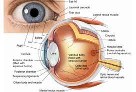
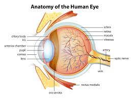
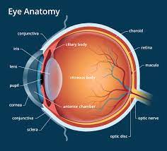
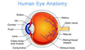
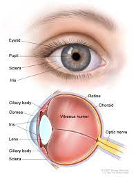
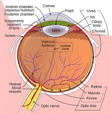
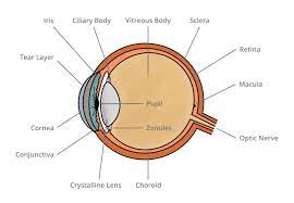
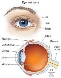
a) Cornea: The cornea is the transparent, dome-shaped layer covering the front of the eye. It acts as a protective barrier and helps to focus incoming light. It is also responsible for most of the eye’s refractive power.
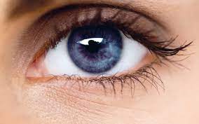
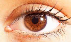
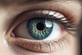
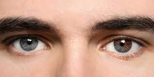
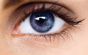
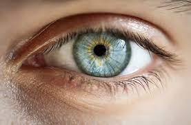
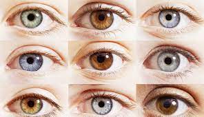
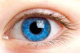
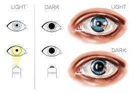
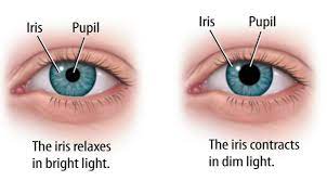
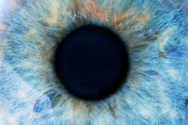
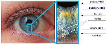
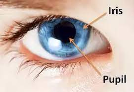
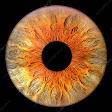
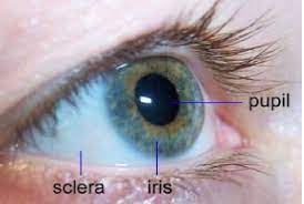
b) Iris and Pupil: The iris is the colored part of the eye surrounding the pupil, which is the black central opening. The iris controls the amount of light entering the eye by adjusting the size of the pupil. The pupil dilates in low light conditions to allow more light in, and constricts in bright light to reduce the amount of light entering the eye.
c) Lens: Behind the iris and pupil lies the crystalline lens. The lens is responsible for further focusing the incoming light onto the retina. It changes shape through a process called accommodation to adjust focus for near and far objects.
d) Retina: The retina is a thin, delicate layer of tissue located at the back of the eye. It contains millions of specialized cells called photoreceptors that convert light into electrical signals. The two types of photoreceptors are rods, which enable vision in low light conditions and detect motion, and cones, which enable us to perceive colors and details in bright light.
e) Optic Nerve: The optic nerve is a bundle of nerve fibers that carries visual information from the retina to the brain, where it is processed and interpreted. The optic nerve is responsible for transmitting the electrical signals generated by the photoreceptors to the visual cortex.
f) Macula and Fovea: The macula is a small area in the center of the retina that is responsible for central vision and visual acuity. Within the macula, there is a tiny depression called the fovea, which contains the highest concentration of cones and provides the sharpest vision.
g) Sclera and Choroid: The sclera is the tough, white outer layer of the eye that maintains its shape and protects the inner structures. The choroid is a layer located between the sclera and the retina, and it supplies oxygen and nutrients to the retina.
-
The Visual Process: Now that we have familiarized ourselves with the eye’s structure, let’s explore how it works to enable vision:
a) Light Refraction: When light enters the eye through the cornea, it is refracted, or bent, by the cornea and the lens to focus it onto the retina. This process ensures that the light rays converge at a precise point on the retina for optimal vision.
b) Photoreceptor Activation: When light reaches the retina, it stimulates the photoreceptors (rods and cones), triggering a chemical reaction that generates electrical signals. Rods are responsible for vision in low light conditions, while cones enable us to perceive colors and details in bright light.
c) Signal Transmission: Once the photoreceptors are activated, they send electrical signals to neighboring cells, called bipolar cells, which in turn transmit the signals to the ganglion cells. The ganglion cells collect and process the signals before sending them out of the eye through the optic nerve.
d) Visual Processing in the Brain: The optic nerve carries the electrical signals to the brain’s visual cortex, located at the back of the brain. Here, the signals undergo extensive processing and interpretation, allowing us to perceive the images and objects in our field of view.
Depth perception, another remarkable feature of human vision, enables us to perceive the relative distances between objects. It is primarily achieved through a combination of binocular cues (using both eyes) and monocular cues (using one eye).
a) Myopia (Nearsightedness): Myopia occurs when the eyeball is too long or the cornea is too curved, causing distant objects to appear blurry. Myopia can often be corrected with glasses, contact lenses, or refractive surgery.
b) Hyperopia (Farsightedness): Hyperopia is the opposite of myopia, where the eyeball is too short or the cornea is too flat, making nearby objects appear blurred. Hyperopia can also be corrected with glasses, contact lenses, or refractive surgery.
c) Astigmatism: Astigmatism results from an irregularly shaped cornea or lens, causing distorted and blurry vision at all distances. It can be corrected with glasses, contact lenses, or refractive surgery.
d) Presbyopia: Presbyopia is an age-related condition that affects the eye’s ability to focus on near objects. It occurs due to the natural hardening of the lens with age. Reading glasses or multifocal lenses are commonly used to correct presbyopia.
e) Color Blindness: Color blindness is a condition where individuals have difficulty distinguishing certain colors, most commonly red and green. It is typically an inherited condition caused by abnormalities in the cones’ pigments in the retina.
f) Cataracts: Cataracts occur when the lens of the eye becomes cloudy, leading to blurred or hazy vision. Cataracts are often age-related but can also be caused by trauma, medications, or certain medical conditions. Surgical removal of the cataract and replacement with an artificial lens is the most common treatment.
Conclusion: Our eyes are truly wonders of nature, allowing us to experience the world in all its glory. From the complex interplay of light refraction to the intricate neural processes in our brain, human vision is a remarkable system that enables us to perceive the beauty and diversity of our surroundings. Understanding how our eyes work not only deepens our appreciation for this extraordinary sense but also highlights the importance of caring for our vision through regular eye exams, adopting healthy eye habits, and seeking appropriate treatments for vision problems. So, let us treasure and marvel at the gift of sight and all that it brings into our lives.


